Groundbreaking Mini-Brain Models Offer New Hope for Personalized Alzheimer’s Treatment and Diagnosis

Scientists at Johns Hopkins Medicine have unveiled groundbreaking research utilizing miniature, lab-grown brain tissues, known as organoids, derived from individuals with Alzheimer’s disease. This pioneering work offers compelling new evidence that these patient-specific brain models can significantly enhance the prediction of how different individuals might respond to medications used to manage the complex psychiatric symptoms associated with Alzheimer’s. The findings, published in the esteemed Alzheimer’s & Dementia: The Journal of the Alzheimer’s Association, represent a critical step forward in the quest for more precise and effective treatments for the millions affected by this devastating neurodegenerative condition.
Alzheimer’s disease, the most prevalent form of dementia, currently impacts over 7 million Americans, with global figures reaching into the tens of millions. The disease not only erodes cognitive function, memory, and the ability to perform daily tasks, but it also frequently triggers a spectrum of neuropsychiatric symptoms. These can include profound anxiety, persistent depression, agitation, apathy, and even psychosis, significantly impacting the quality of life for patients and placing immense strain on caregivers. While there is no cure for Alzheimer’s, treatments like selective serotonin reuptake inhibitors (SSRIs) are commonly prescribed to alleviate these challenging symptoms. However, the efficacy of these medications varies dramatically from person to person, leaving a critical unmet need for personalized therapeutic strategies.
The Johns Hopkins team’s research focused on hindbrain organoids, miniature models designed to mimic a specific region of the brain crucial for regulating fundamental life functions such as breathing, sleep, and heart rate. By studying these organoids, researchers aimed to identify molecular signatures that could predict a patient’s response to escitalopram oxalate, a widely used SSRI. This approach holds the potential to move beyond a one-size-fits-all treatment model, ushering in an era of precision medicine for Alzheimer’s disease.
From Blood Cells to Brain Organoids: A Technological Leap
The genesis of this research lies in a meticulous process of cellular reprogramming. Researchers at the NIH-funded Johns Hopkins Alzheimer’s Disease Research Center obtained blood samples, with explicit consent, from individuals diagnosed with Alzheimer’s disease, as well as from healthy control participants. These blood cells were then coaxed back into a pluripotent stem cell-like state – a remarkable feat of biotechnology that transforms specialized cells into cells with the potential to develop into any cell type in the body. These reprogrammed cells, known as induced pluripotent stem cells (iPSCs), served as the foundational material for generating the brain organoids.
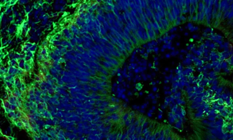
Using iPSCs from both Alzheimer’s patients and healthy individuals, the researchers cultivated hindbrain organoids. These miniature brain models were engineered to contain specialized brain cells, or neurons, that produce serotonin, a key neurotransmitter implicated in mood regulation and targeted by SSRI medications. Through carefully controlled laboratory conditions, these iPSCs were guided to self-organize into small, pea-sized clusters of brain tissue that closely resemble the architectural and cellular organization of the hindbrain. The study’s scale is noteworthy; it encompassed hundreds of organoids, each representing an individual patient’s genetic makeup and disease profile, alongside those from healthy individuals. Dr. Vasiliki Machairaki, the study’s lead investigator and an associate professor of genetic medicine at Johns Hopkins University School of Medicine, described it as potentially one of the most extensive brain organoid studies conducted to date in the field of Alzheimer’s research.
Unveiling Molecular Hallmarks of Alzheimer’s
Upon analysis, the patient-derived organoids exhibited distinct molecular characteristics that mirrored key biological processes observed in Alzheimer’s disease. Compared to organoids derived from healthy individuals, those grown from the cells of Alzheimer’s patients displayed significant differences in the expression of proteins involved in critical brain functions. These included proteins essential for neuronal communication, inflammatory pathways, and other molecular mechanisms known to be dysregulated in Alzheimer’s.
The critical juncture of the study involved treating these organoids with escitalopram oxalate. The researchers observed varied responses: in some Alzheimer’s patient-derived organoids, the medication led to an increase in proteins associated with serotonin signaling and inter-neuronal communication – pathways that are the intended targets of antidepressant medications. Conversely, other organoids showed minimal or no discernible molecular alteration in response to the drug. This differential response is precisely what the researchers sought to uncover, as it points towards the possibility of predicting treatment efficacy based on an individual’s unique molecular profile.
"We used these organoids to model how some patients’ tissue may respond to a commonly prescribed SSRI," explained Dr. Machairaki. "On a large-scale level, our model may eventually be used to identify subgroups of patients, based on underlying molecular mechanisms, who are more likely to respond to certain drugs and thus help us to create precise, targeted treatments in the long run." This statement underscores the transformative potential of the research, moving towards a future where treatments can be tailored to the specific biological underpinnings of an individual’s Alzheimer’s disease.
Extracellular Vesicles: Tiny Messengers with Big Implications
Beyond observing molecular changes within the organoids themselves, the research team delved into the role of extracellular vesicles (EVs). These are minute particles released by cells, acting as tiny envelopes that carry cellular information, including proteins and genetic material, to other cells. The scientists hypothesized that EVs released by Alzheimer’s organoids might serve as valuable biomarkers for diagnosing the disease, gauging its progression, and even predicting treatment responses.
Before and after the escitalopram treatment, the researchers meticulously examined the protein content of EVs released by both patient-derived and healthy control organoids. They discovered that these vesicles contained proteins critical for various brain functions, including neuronal communication, memory formation, and the release of neurotransmitters.
Significantly, EVs from organoids derived from individuals with Alzheimer’s disease exhibited discernible alterations in several disease-associated proteins. Notably, the levels of proteins such as RAB3A, NSF, and ATCAY were found to be lower in these Alzheimer’s EVs. These proteins are vital for the normal signaling processes between brain cells.
Following escitalopram treatment, specific protein levels within the EVs showed an increase in certain samples. These changes were particularly pronounced in proteins linked to serotonin signaling and synaptic pathways – the very pathways that SSRIs are designed to influence. The observed variation in EV protein profiles, mirroring the cellular response of the organoids, further strengthens the hypothesis that these tiny vesicles could hold the key to predicting drug response.
"Some organoids displayed strong molecular responses, while others showed little or no change," stated Dr. Machairaki. "This variation raises the possibility that extracellular vesicles from brain organoids could eventually help identify which patients are most likely to benefit from a particular treatment." This finding opens up exciting avenues for developing non-invasive diagnostic and prognostic tools.
The Road Ahead: Enhancing Realism and Clinical Application
The current study represents a significant foundational achievement, but Dr. Machairaki and her team are already looking towards the future, with plans to develop even more sophisticated organoid models. Their vision includes incorporating other crucial cell types, such as immune cells, and engineering vascular-like networks that mimic the intricate blood vessel systems found in the human brain. The inclusion of these elements would imbue the organoids with a greater degree of realism, making them even more representative of living human brain tissue.
With continued research and refinement, the hope is that extracellular vesicles, isolated from easily accessible bodily fluids like blood, could eventually function as a form of "liquid biopsy." Such a test would revolutionize Alzheimer’s diagnostics, potentially enabling early detection, accurate staging of disease progression, and precise identification of a patient’s specific disease subtype. This personalized approach to diagnosis could then inform tailored treatment strategies.
"Our study suggests that large-scale, patient-derived brain organoids and the vesicles they secrete can help us stage Alzheimer’s disease, investigate the mechanisms that drive it and assess how patient subgroups may respond to different treatments," Dr. Machairaki reiterated. The implications of this research extend beyond drug response prediction; it also offers a powerful platform for unraveling the complex molecular mechanisms underlying Alzheimer’s disease itself, a critical endeavor for the development of novel therapeutic targets.
The study was partially funded by the National Institutes of Health (NIH), underscoring the federal government’s commitment to advancing Alzheimer’s research. The collaborative effort involved a multidisciplinary team of scientists from Johns Hopkins University, Tymora Analytical Operations, and the University of Rochester School of Medicine and Dentistry. This collaborative spirit is often essential in tackling complex diseases like Alzheimer’s.
The development of patient-derived organoids represents a significant advancement in modeling neurological disorders. Historically, research has relied on animal models or post-mortem human brain tissue, which often have limitations in fully recapitulating the dynamic and complex nature of human brain diseases. Organoids, by contrast, offer a living, dynamic, and patient-specific platform for studying disease processes and testing therapeutic interventions in a controlled laboratory setting. This research from Johns Hopkins Medicine is a testament to the power of cutting-edge biotechnology in illuminating the complexities of Alzheimer’s disease and paving the way for a future where personalized care is not just an aspiration, but a reality for patients.
Related posts: