
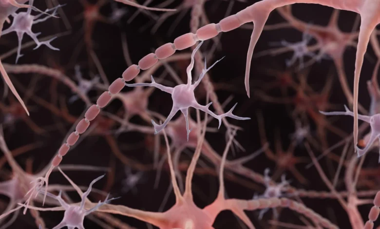
This discovery, which bridges the fields of cell biology, biophysics, and neuroscience, challenges the foundational assumptions of how neural circuits operate. The implications extend from basic human physiology to the potential for new diagnostic frameworks in neurodegenerative medicine.
A Paradigm Shift in Neural Anatomy
The conventional understanding of the neuron, as taught in undergraduate biology curricula worldwide, posits that the axon serves as a passive transmission line. While scientists have long recognized "varicosities"—bulges along the axon—these were almost exclusively categorized as synaptic terminals, the specialized sites where neurotransmitters are released to signal adjacent cells. In pathological conditions, such as Parkinson’s disease or traumatic brain injury, larger, irregular beading is often observed as a hallmark of cellular distress or structural degradation.
The research published in Nature Neuroscience on December 2, 2024, by Shigeki Watanabe and his team, introduces a critical distinction: "non-synaptic varicosities." By utilizing high-pressure freezing electron microscopy, the researchers successfully captured the native state of axonal architecture in mouse neurons, avoiding the structural artifacts often introduced by traditional chemical fixation and dehydration. The result was a high-resolution revelation of repeating, nanoscale pearls that exist in perfectly healthy, functional axons. These structures are not signs of decay, but rather a standard, dynamic feature of neuronal design.
Chronology of the Discovery
The path to this discovery was iterative, spanning over a decade of observations across different biological models. The investigation began with Watanabe’s curiosity regarding repeating axonal patterns in C. elegans (nematodes), which prompted a collaboration with Swiss scientist Graham Knott.
- 2012: A Harvard University study first documented periodic "skeletal" components within axons, providing a potential structural basis for axonal organization.
- 2023–2024: The Johns Hopkins team, led by graduate student Jacqueline Griswold, tested whether these skeletal proteins were responsible for the pearling. When the removal of these components failed to eliminate the pearls, the team shifted their focus toward membrane biophysics.
- December 2, 2024: Publication of the primary study in Nature Neuroscience, confirming that pearling is a widespread, non-pathological feature in mouse models.
- November 2025 – February 2026: Researchers, including Chelsy R. Eddings and Shigeki Watanabe, extended the findings to human cortical tissue obtained during epilepsy surgeries, demonstrating that the phenomenon is not limited to mice.
- July 2026: A study published in PLOS Biology by researchers at the University of Edinburgh utilized zebrafish to demonstrate that chemical signaling pathways can actively modulate axon diameter, providing a secondary layer of evidence that axon shape is a regulated variable rather than a static constant.
The Physics of Signaling: Membrane Tension and Geometry
To understand why axons adopt this beaded morphology, the Johns Hopkins team partnered with theoretical biophysicist Padmini Rangamani. Their mathematical modeling suggests that the pearling is a consequence of mechanical forces and membrane dynamics rather than a rigid internal scaffold.
The research revealed that the membrane’s physical properties—specifically its stiffness and the tension exerted by the surrounding environment—dictate the size and frequency of the pearls. By manipulating the concentration of cholesterol within the axonal membrane, the team was able to alter the degree of "pearling." A critical finding here is the impact on signal transmission: the width of the axonal segment determines the resistance encountered by ions as they travel along the axon.
"A wider space in the axons allows ions to pass through more quickly and avoid traffic jams," Watanabe noted in the initial 2024 announcement. This suggests that the brain may dynamically adjust the "pearl" architecture to tune the speed of communication between neurons. When the researchers stimulated the neurons with high-frequency electrical pulses, they observed the pearls physically enlarging—an effect that persisted for at least 30 minutes and correlated with a measurable slowing of electrical signal velocity. This plasticity indicates that the axon is an active participant in signal processing, capable of reshaping itself in real-time in response to activity.
Clinical Implications and Future Directions
The shift from viewing axons as static cables to seeing them as dynamic, modulated structures carries profound implications for clinical neurology. For years, the presence of axonal beading in diagnostic imaging or histology has been treated as an unequivocal indicator of neurodegeneration or cell death. With the recognition that healthy axons possess a form of nanoscale pearling, the medical community must now recalibrate its diagnostic criteria to distinguish between physiological architecture and pathological damage.
The work by Eddings et al., published in Neuron, further bridges the gap by linking these membrane changes to "ultrafast endocytosis," a process essential for recycling the neuronal membrane after chemical signaling. This suggests that the pearled regions may serve as strategic reservoirs for membrane components, allowing the neuron to maintain rapid-fire communication without exhausting its surface area.
Broader Impact and Scientific Consensus
The National Institute of Mental Health (NIMH) has recognized the significance of this work by awarding a Multiple Principal Investigator grant to Watanabe and Rangamani. The ongoing project seeks to create a comprehensive computational model of axonal signaling, integrating these physical properties into our broader understanding of brain function.
The research also opens new avenues for pharmaceutical intervention. If axon diameter and pearling can be influenced by specific chemical signaling pathways—as evidenced by the Edinburgh zebrafish study—it is theoretically possible that future therapies could target these mechanisms to enhance neural connectivity in patients with neurodegenerative conditions or to restore function in damaged neural circuits.
However, researchers remain cautious about over-extrapolating the findings. While pearled axons have now been identified in human tissue, the extent of this architecture’s variability across different brain regions and age groups remains to be mapped. The current findings represent a "first-look" at a level of structural complexity that was previously invisible to standard light microscopy.
As the scientific community moves forward, the "pearled axon" model serves as a reminder of the limitations of historical imaging techniques. By shifting from the standard paradigm of fixed, static anatomy to a fluid, biophysical model, researchers are uncovering a layer of neural regulation that may be fundamental to how the brain encodes information. The collaboration between biologists and physicists in this space underscores a growing trend in neuroscience: the realization that biological function cannot be understood in isolation from the underlying physics of cellular structure.
The transition from a "smooth tube" theory to a "beaded architecture" model is more than a semantic or illustrative change; it is an invitation to re-examine the mechanics of thought itself. As laboratory techniques like high-pressure freezing continue to provide clearer windows into the nanoscale environment of the brain, the next decade of neuroscience research will likely focus on how these microscopic pearls facilitate the complex, high-speed computations that define human cognition, memory, and behavior. The textbook illustration of the neuron may have been updated, but the true depth of its functional significance is only beginning to be understood.
Related posts: