The Anatomy of a Misunderstanding
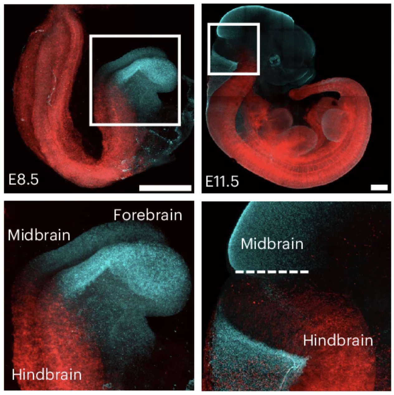
The confusion appears to have originated from a study highlighting how specific genetic markers differentiate the hindbrain from the midbrain and forebrain during the earliest stages of embryonic growth. By utilizing fluorescent protein tagging in mice, researchers demonstrated that cells destined to become the hindbrain are distinct from those designated for the rest of the brain from an incredibly early developmental checkpoint.
The media coverage seized upon the phrase "two separate brains" to describe these distinct cellular lineages. However, in the context of developmental biology, this is a categorical error. A "brain" is defined by its integrated function, its physical connectivity, and its role as a centralized hub for the nervous system. The researchers were not describing two functioning, autonomous brains, but rather the establishment of early regional identities within a single developing neural tube.
Chronology of Development: From Sheet to Tube
To understand why these claims are misleading, one must look at the timeline of human neurodevelopment. The process begins with gastrulation, where the embryo organizes itself into three primary germ layers: the endoderm, the mesoderm, and the ectoderm. The nervous system originates from the ectoderm.
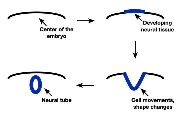
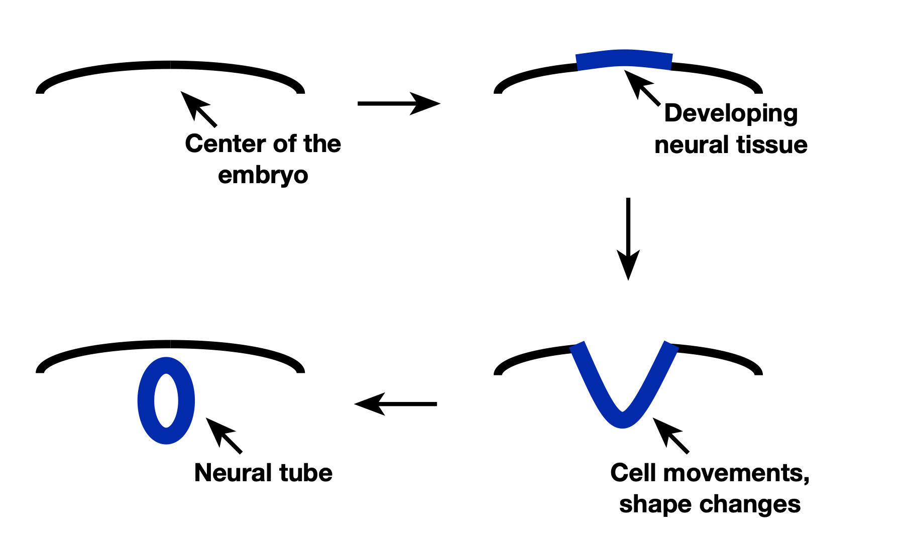
- The Neural Plate Phase: A central band of the ectoderm thickens to form the neural plate. This structure is essentially a flat, primitive sheet of precursor cells.
- Neurulation: The edges of the neural plate fold upward and inward, eventually fusing to form the neural tube. This tube is the structural precursor to the entire central nervous system, including the brain and spinal cord.
- Regionalization: Even as the tube forms, it begins to "know" its orientation—head to tail and back to belly. The new research focuses on this specific stage, showing that cells are assigned their regional identity (forebrain, midbrain, or hindbrain) during or even slightly before the tube completes its closure.
The findings suggest that the "decision" for a cell to become part of the hindbrain versus the forebrain is made earlier than previously assumed, effectively partitioning the developing brain into territories before the organs fully manifest.
Decoding the Genetic Blueprint
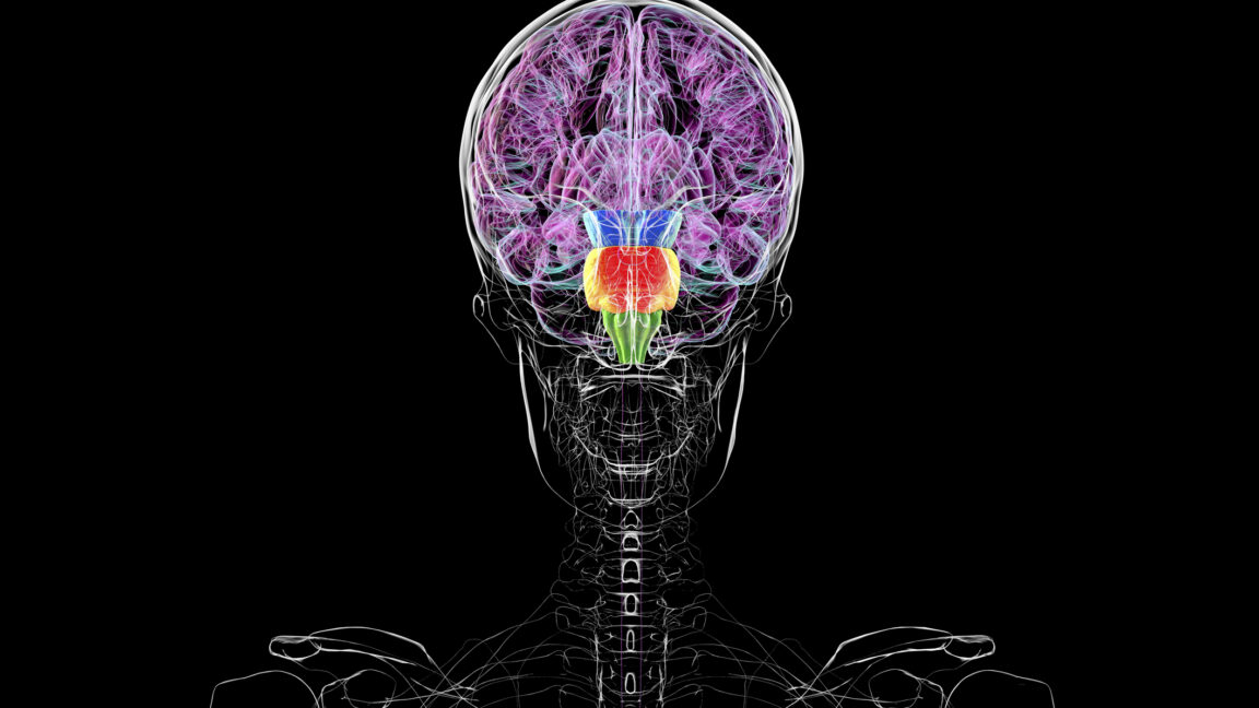
The research team employed advanced genetic engineering to visualize this process. By modifying genes that regulate early brain development to express fluorescent proteins, they created "glowing" maps of the embryo. The hindbrain emerged in a distinct red hue, while the forebrain and midbrain were marked in cyan.
The data revealed a striking degree of fidelity: roughly 96 percent of descendant cells from a single early precursor remained within their assigned segment. This indicates that once a group of cells is "assigned" to the hindbrain, they rarely migrate to or adopt the identity of the midbrain. This is not the creation of a second brain, but rather the execution of a highly choreographed spatial program that ensures the brain has the correct proportions and specialized regions.
Implications for Developmental Biology
The implications of this study are profound for fields beyond basic anatomy, particularly in regenerative medicine and stem cell research. For decades, scientists have sought to understand how to nudge pluripotent stem cells into becoming specific types of neurons. If we know that these cells require specific signaling environments to adopt a "hindbrain identity" versus a "forebrain identity," we can better design protocols to grow organoids—miniature, lab-grown brain models—that accurately mimic human development.
The study also clarifies how a limited set of signaling molecules, such as the Sonic hedgehog protein or Bone Morphogenetic Proteins (BMPs), can produce such vast complexity. The answer lies in the "developmental history" of the cells. Because a cell "knows" it is part of the midbrain due to its early regionalization, it interprets the same BMP signal differently than a cell in the spinal cord would. This allows the body to recycle signaling pathways to perform different tasks in different tissues.
Expert Perspectives and Scientific Consensus
While the university press release likely intended to emphasize the discovery of these early "fate maps," the resulting public discourse highlights a growing disconnect between complex biological research and popular science communication. Experts in the field have been quick to clarify the terminology.
"We are looking at the foundational zoning laws of the brain," noted one developmental biologist familiar with the research. "To suggest this creates ‘two brains’ is akin to saying that because a city is divided into residential and commercial zoning, it is actually two different cities. It is a single, integrated urban plan, just with different functional sectors."
The consensus remains that the brain is a singular, unified organ. While it is composed of modular structures—the brainstem, the cerebellum, the cortex, and so on—these parts are physically and neurologically interconnected. The "two brains" narrative fails to account for the massive web of axonal pathways that communicate between these regions, ensuring that the organism functions as a coherent whole.
Broader Impact: Why the Distinctions Matter
Understanding these boundaries is essential for identifying the origins of neurodevelopmental disorders. Many conditions, such as microcephaly or various forms of developmental brain malformation, occur when these early signals or regional divisions are disrupted. If the "zoning" process fails, the resulting structure is often compromised, leading to profound functional deficits.
Furthermore, this research reinforces the importance of the environment surrounding the embryo. The signals that inform cells of their position—diffusing from nearby tissues—are delicate. Environmental stressors or chemical exposures that interfere with these early signaling gradients can have disproportionate effects on specific brain regions.
Conclusion: Moving Beyond the Headline
The excitement surrounding this research is, in many ways, justified; it provides a clearer picture than ever before of how the human brain emerges from a simple, uniform layer of cells. However, the interpretation of this as a dual-brain system is a distortion that undermines the elegant reality of biology.
We possess a singular brain, a complex and highly specialized organ that achieves its functionality through precise, early-stage regionalization. The research does not challenge our fundamental understanding of human anatomy; it refines it, providing a more detailed map of the processes that shape the most complex structure in the known universe. By moving past the sensationalism, we gain a deeper appreciation for the intricate, step-by-step assembly required to build the organ that allows us to perceive, think, and exist.
Related posts: