Groundbreaking Stanford Medicine Study Reveals Potential to Reverse Osteoarthritis by Targeting Aging Proteins in Knee Cartilage

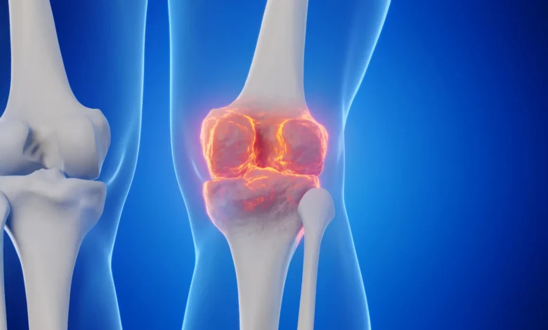
In a development that could fundamentally alter the landscape of orthopedic medicine, researchers at Stanford University have identified a biological pathway capable of regenerating damaged knee cartilage. By inhibiting a specific protein that accumulates with age, the research team successfully reversed natural cartilage degradation in mice and demonstrated the potential to protect joints from the long-term damage typically associated with traumatic injuries, such as ACL tears. The findings, published in the journal Science, suggest that the human body may possess an inherent, latent capacity for joint repair that can be unlocked through pharmacological intervention.
For millions of individuals worldwide, the diagnosis of osteoarthritis represents a slow, painful trajectory toward total joint replacement. Unlike other tissues in the body that exhibit some level of regenerative capacity, articular cartilage—the smooth, slippery tissue covering the ends of bones—has long been considered incapable of self-repair. The current standard of care is largely palliative, focusing on symptom management, physical therapy, and, ultimately, invasive surgical procedures. The Stanford study introduces a paradigm shift, proposing that the breakdown of cartilage is not an irreversible process but rather a consequence of an aging-related signaling pathway that can be modulated.
The Role of 15-PGDH: A Gerozyme in Action
The research centers on an enzyme known as 15-PGDH (15-hydroxyprostaglandin dehydrogenase). Scientists have classified this protein as a "gerozyme"—a term describing enzymes that increase in concentration as an organism ages, contributing to the progressive decline of tissue health. Previous studies led by Helen Blau, PhD, director of the Baxter Laboratory for Stem Cell Biology, established that 15-PGDH acts as a master regulator of tissue aging. By suppressing this protein, the research team was able to increase muscle mass and endurance in older mice, effectively "rejuvenating" the tissue.
The transition from muscle studies to cartilage research was motivated by a critical question: if 15-PGDH regulates cellular aging in muscle and bone, does it play a similar role in the deterioration of articular cartilage? To investigate, the team measured levels of 15-PGDH in the knee cartilage of both young and aging mice. The results showed a significant correlation between age and protein expression; in older animals, the levels of the gerozyme were roughly double those found in younger subjects.
Mechanisms of Cellular Rejuvenation
The most surprising discovery during the investigation was the mechanism of regeneration. Historically, researchers looking for ways to treat cartilage damage have focused on identifying and stimulating stem cells within the joint. However, the Stanford team found that no such stem cell recruitment was necessary. Instead, the treatment caused existing cartilage cells, known as chondrocytes, to alter their gene expression patterns, shifting from a state of degradation to one of active repair and youthful function.
The treatment process involved a small-molecule inhibitor designed to block 15-PGDH. When administered to older mice, either through systemic abdominal injection or direct intra-articular delivery, the effect was immediate and pronounced. The cartilage, which had been thin and structurally compromised, thickened across the surface of the joint. Crucially, the cells began producing healthy hyaline cartilage—the specialized tissue required for smooth joint articulation—rather than fibrocartilage, which is generally less durable and less effective at reducing friction.
Clinical Implications for ACL Injuries and Beyond
Traumatic knee injuries, particularly anterior cruciate ligament (ACL) tears, are a major precursor to early-onset osteoarthritis. Despite surgical repair of the ligament, approximately half of all patients experience post-traumatic osteoarthritis within 15 years of their initial injury. This suggests that the initial trauma initiates a cascade of molecular signaling that the current standard of care fails to address.
In a controlled experiment, the researchers administered the 15-PGDH inhibitor twice weekly for four weeks following a simulated ACL injury in mice. The results were stark: the treatment group exhibited a significantly lower incidence of osteoarthritis compared to control groups. Treated mice displayed improved mobility and greater weight-bearing capacity on the injured limb, indicating that the molecular intervention successfully mitigated the secondary degenerative effects of the injury.
Human Tissue Validation and Future Trials
To assess the translational potential of these findings, the team conducted experiments on human cartilage samples obtained from patients undergoing total knee replacement surgery. After one week of exposure to the 15-PGDH inhibitor, the tissue showed a significant reduction in the presence of 15-PGDH. Simultaneously, there was a measurable decrease in the expression of genes associated with inflammation and cartilage breakdown, coupled with an increase in markers for healthy, functional cartilage production.
While these results provide strong evidence for the efficacy of the treatment, the research team emphasizes that this is still a preclinical development. Clinical trials are necessary to determine safety, optimal dosage, and long-term efficacy in human patients. However, the timeline for such trials may be accelerated. Because an oral 15-PGDH inhibitor has already completed Phase 1 clinical trials for muscle weakness, researchers have a foundational safety profile upon which to build.
"Phase 1 clinical trials of a 15-PGDH inhibitor for muscle weakness have shown that it is safe and active in healthy volunteers," Blau noted. "Our hope is that a similar trial will be launched soon to test its effect in cartilage regeneration."
The Economic and Healthcare Burden of Osteoarthritis
The societal cost of failing to address osteoarthritis is immense. In the United States alone, the condition affects one in five adults, representing a massive burden on the healthcare system. Estimates suggest that direct healthcare costs associated with the disease exceed $65 billion annually, a figure that is expected to climb as the global population ages. Currently, the medical community is limited to treating the pain and inflammation associated with the condition until the joint becomes so degraded that replacement surgery is the only remaining option.
A pharmacological approach that can slow, stop, or even reverse the progression of osteoarthritis would represent one of the most significant advancements in orthopedic medicine in the last century. By focusing on the underlying molecular causes of tissue aging rather than merely masking the symptoms, the research led by Blau and Nidhi Bhutani, PhD, offers a glimpse into a future where joint replacement might be avoided entirely.
Scientific Context and Ethical Disclosures
The study, which was supported by the National Institutes of Health and various private foundations, involved a collaborative effort between Stanford Medicine and the Sanford Burnham Prebys Medical Discovery Institute. The research highlights a shift in focus toward "geroscience," a field dedicated to understanding the biological mechanisms of aging to treat age-related diseases as a group.
As with any significant medical breakthrough, there are commercial and academic interests at play. The researchers have noted that they are inventors on patent applications held by Stanford University regarding the use of 15-PGDH inhibition for tissue rejuvenation. These patents have been licensed to Epirium Bio, a biotechnology company co-founded by Dr. Blau. These disclosures, standard in high-impact medical research, underscore the transition of the project from basic laboratory discovery to potential commercial development.
Conclusion
The discovery that cartilage cells can be reprogrammed to repair themselves by inhibiting a single protein is a testament to the power of molecular biology. By viewing osteoarthritis not as an inevitable "wear and tear" process, but as a biological phenomenon driven by age-related protein activity, the Stanford team has opened a new frontier in regenerative medicine. If future clinical trials mirror the success observed in animal models and human tissue samples, the medical community may soon have at its disposal an oral or injectable therapy capable of restoring the structural integrity of joints, potentially sparing millions of people from the pain and functional limitations of chronic joint disease.