Unlocking the 3D Genome: A Breakthrough in Alzheimer’s Research Reveals New Targets for Treatment


A collaborative team of researchers from Carnegie Mellon University, the University of Pittsburgh, and the University of Washington has unveiled a sophisticated new layer of biological complexity underlying Alzheimer’s disease, identifying significant disruptions in the three-dimensional organization of the genome within brain cells. This study, published in the journal Science, moves beyond the traditional focus on amyloid-beta plaques and tau tangles, suggesting that the structural "folding" of DNA plays a critical role in the molecular pathology of the disease. By integrating single-cell technology with advanced artificial intelligence, scientists have provided a roadmap for potential future therapeutic interventions.
The Evolution of Alzheimer’s Diagnostics and Research
For decades, the medical community’s understanding of Alzheimer’s disease has been dominated by the "amyloid cascade hypothesis," which posits that the accumulation of amyloid-beta proteins and the subsequent formation of neurofibrillary tangles (tau) are the primary drivers of cognitive decline. While these features are undeniable hallmarks of the disease, clinical trials targeting these proteins have yielded mixed results, leaving a significant portion of the pathology unexplained.
With Alzheimer’s currently affecting approximately seven million Americans—a figure projected to nearly double by 2050 as the population ages—the search for novel therapeutic targets has become a critical public health priority. The current research marks a departure from traditional models by examining the physical state of the genome itself. DNA, when stretched out, measures approximately two meters in length; inside the cell nucleus, it is coiled into complex, three-dimensional structures. This organization is not merely structural; it is functional. The way DNA folds dictates which genes are accessible to the cellular machinery, effectively serving as an architectural regulator of gene expression.
Integrating Multi-Omics and Artificial Intelligence
To achieve this level of granular detail, the research team utilized a multi-disciplinary approach. The study involved analyzing postmortem samples from the prefrontal cortex of individuals who participated in long-term longitudinal dementia studies. By employing a technique known as GAGE-seq, the researchers were able to measure gene expression and the 3D contact patterns of the genome simultaneously within individual cells.
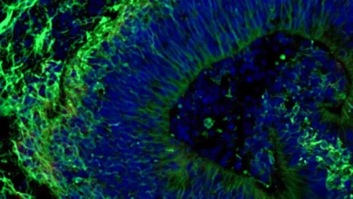
This data was then synthesized with spatial transcriptomic mapping, which allowed the team to visualize precisely where these molecular shifts occurred within the intact architecture of brain tissue. A cornerstone of the project was the development of "Hicformer," a specialized deep learning model. Hicformer acts as a computational bridge, inputting DNA sequence information alongside patterns of genome folding to predict how specific cell types will behave under various conditions.
Xinyue Lu, a doctoral student in Computational Biology and co-lead of the research, noted that the model serves as a "computational test bed." This allows scientists to simulate how alterations in the 3D structure of the genome translate into changes in gene activity, providing a high-speed way to screen for mechanisms that might be fueling disease progression.
Defining the Structural Collapse: "Compartment Mingling"
The research revealed that in the brain cells of individuals with Alzheimer’s disease, the genome’s architectural integrity is significantly compromised. Under normal, healthy conditions, the genome is organized into distinct "compartments"—specific regions that are either active or inactive. These boundaries keep the cell’s regulatory systems functioning in an orderly fashion.
In the Alzheimer’s-affected brain, however, these boundaries appear to break down. The researchers identified a phenomenon termed "increased compartment mingling," where the distinct separation between active and inactive chromatin—the protein-DNA complex that packages the genome—becomes blurred. This leads to a cascade of functional consequences:
- Reduced Gene Activity: Cells exhibiting high levels of compartment mingling showed a marked decrease in overall gene expression.
- Regulatory Disconnection: Interactions between genes and the regulatory elements that "switch" them on or off were found to be weakened, disrupting cellular stability.
- Synaptic Dysfunction: The structural reorganization was specifically linked to the downregulation of gene programs essential for neuronal and synaptic health.
- Microglial Senescence: In microglia—the brain’s primary immune cells—the researchers observed links to senescence-related programs, suggesting that the architectural changes may impede the immune system’s ability to clear debris and manage inflammation.
Chronology of the Discovery
The road to these findings was a years-long effort requiring the synchronization of high-throughput biology and computational power. The process began with the procurement of high-quality postmortem brain tissue from longitudinal studies, ensuring the samples were matched for age and clinical history.
In the early stages, the team focused on developing the necessary spatial transcriptomic pipelines to preserve the "tissue context" of the samples. Once the GAGE-seq data was collected, the team spent significant time training the Hicformer model to recognize the signatures of genome folding. By late 2023 and early 2024, the integration of these datasets revealed the consistent signature of 3D reorganization across multiple brain cell types. This culminated in the peer-review process and the final publication in Science, which sets the stage for a new generation of studies focused on genomic structural stability.
Broader Impact and Clinical Implications
The findings represent a fundamental shift in how the medical research community approaches Alzheimer’s pathology. By establishing 3D chromatin alterations as a key component of the disease, the researchers have effectively expanded the "druggable" genome.
"Alzheimer’s disease cannot be understood one layer at a time," said Jian Ma, the Ray and Stephanie Lane Professor of Computational Biology and the study’s supervisor. "By integrating genome folding, cell state, and tissue context, we can move beyond cataloging disease-associated changes toward understanding how they fit together and which mechanisms to test next."
The implications for drug development are significant. If the breakdown of 3D genome structure contributes to the suppression of vital synaptic genes, then therapies aimed at stabilizing the chromatin—or restoring the boundaries between active and inactive genome compartments—could theoretically mitigate the cognitive decline associated with the disease. Furthermore, the ability to pinpoint these changes in microglia offers a potential path to modulating the brain’s inflammatory response, a factor long known to exacerbate the symptoms of dementia.
Expert Perspectives and Future Directions
Hansruedi Mathys, an assistant professor of neurobiology at the University of Pittsburgh who directed the Pitt arm of the study, emphasized the gravity of the results. "We know the classic hallmarks, but our results establish higher-order chromatin alterations as a component of the molecular pathology associated with the disease," he stated.
The research was made possible through the support of the National Institutes of Health, reflecting the high priority placed on uncovering the underlying mechanics of neurodegeneration. While the current study is a foundational leap forward, the authors acknowledge that the next phase of research will require rigorous testing. The team plans to focus on specific regulatory regions identified by Hicformer to determine if they can be targeted with small-molecule drugs or gene-editing technologies to rescue cells from the degenerative process.
As the scientific community digests these findings, the focus will likely shift toward the "spatial" aspect of the disease. Understanding why these genomic shifts occur in specific regions of the prefrontal cortex—and whether similar disruptions are present in the hippocampus or other areas early in the disease trajectory—will be essential for developing diagnostic tools that can detect Alzheimer’s before significant tissue loss occurs.
This study does not render the amyloid-tau hypothesis obsolete; rather, it contextualizes it within a much broader, more dynamic framework of genomic health. By looking at the cell as an integrated, three-dimensional system, researchers are moving closer to a holistic understanding of the disease, one that may finally provide the breakthroughs necessary to change the trajectory of this devastating condition for millions of patients worldwide.