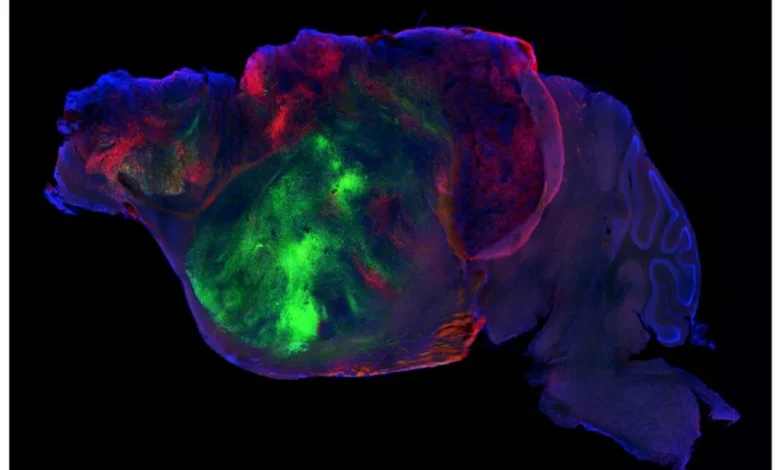
In a landmark experiment published in the journal Nature, a research team at Stanford University has successfully replaced a significant portion of a developing mouse brain with human cortical organoids. This achievement marks a new chapter in regenerative medicine and neurobiology, offering a sophisticated, albeit experimental, model for studying human-specific brain development and the complex pathologies of neurological disorders. By genetically engineering mice to lack a native cortex and filling the void with human-derived tissue, scientists have created a chimeric model that bridges the long-standing gap between in vitro organoid studies and the intricate environment of a living organism.
The limitations of traditional "organoids"—three-dimensional clusters of cells grown in laboratory dishes—have long frustrated neuroscientists. While these structures mimic the basic cellular architecture of human organs, they exist in a vacuum, disconnected from the systemic pressures of blood flow, endocrine signaling, and the complex, long-range neural connectivity that defines human cognition. The Stanford study, led by researchers in the lab of Dr. Sergiu Pasca, addresses these deficiencies by providing the human tissue with an active biological host environment.
A Chronology of Developmental Engineering
The methodology behind this study was as meticulous as it was radical. The researchers identified a specific gene essential for the chromosomal separation process during cell division. By targeting this gene to be silenced within the developing mouse cortex, they were able to halt the formation of the mouse’s own cortical structures. This process, while seemingly destructive, resulted in a mouse that could survive the gestation period, provided the researchers implemented strict post-natal care protocols.
To ensure the survival of these cortex-deficient subjects, the team culled litters to reduce competition for nursing and provided the remaining pups with high-calorie nutritional supplements. This intervention allowed the mice to bypass the immediate lethal effects of a missing cortex, creating a "biological vacuum" waiting to be filled. Into this space, the team implanted human neural stem cells derived from human induced pluripotent stem cells (iPSCs).
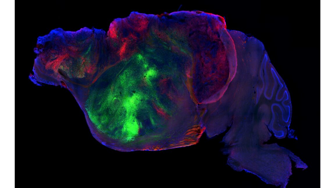
The integration was remarkably successful. Approximately 85 percent of the implanted mice showed successful graft integration. Within these hosts, the human cells proliferated to account for nearly 92 percent of the cortical volume. The human cells did not merely exist in a dormant state; they matured, formed diverse neuronal types, and extended axonal projections that reached as far as the spinal cord, suggesting a functional integration into the mouse’s existing nervous system.
Data and Behavioral Analysis
The primary challenge for the researchers was to determine whether this "humanized" cortex provided any tangible benefit to the host beyond structural filling. To quantify this, the team employed a multi-modal approach, combining machine-learning-driven behavioral analysis with standard neuro-behavioral testing.
When placed in a controlled environment and monitored via high-resolution video, the mice with the humanized cortex exhibited behavioral patterns distinct from both normal mice and those with complete cortical ablation. In body weight comparisons, the humanized mice demonstrated an intermediate phenotype, suggesting that while the human tissue provided some restorative function, it did not reach the metabolic or physiological benchmarks of a fully native mouse cortex.
Memory and motor coordination tests provided further nuance. In standard maze navigation, mice lacking a cortex performed at the level of random chance. The humanized cohort, however, demonstrated measurable improvement, performing above chance, though still failing to match the proficiency of control subjects. Intriguingly, in associative memory tasks—a complex function of the cortex—the humanized mice showed no significant improvement over the cortex-deficient control group. This discrepancy suggests that while the human cells are capable of forming basic connections, they may lack the organized, high-level circuitry required for complex cognitive tasks.
The Problem of Structural Organization
Despite the successful integration of the human cells, the study revealed a significant hurdle: the lack of laminar organization. In a naturally developed human brain, cortical neurons arrange themselves in specific, functional layers. In these mouse models, while specific cell types remained in close proximity, the characteristic layered structure was notably absent.
This observation highlights a fundamental tension in neurobiology: the difference between cell survival and functional circuit assembly. The human cells appear to be following an intrinsic genetic program that allows them to differentiate and survive, but they are clearly missing the spatial cues from the mouse environment necessary to organize into a fully functional, layered cortex. Whether this lack of structure is a result of the mouse’s brain chemistry failing to communicate with human cells, or simply a byproduct of the implantation process, remains a subject of intense investigation.
Scientific Implications and Future Outlook
The broader implications of this work are significant for the study of neurodevelopmental and psychiatric diseases, such as autism, schizophrenia, and ALS. Currently, these diseases are difficult to study because they involve human-specific interactions that mouse models cannot replicate. By creating a "humanized" environment, researchers may eventually be able to observe how disease-specific mutations manifest within a living, albeit chimeric, system.

However, the scientific community remains cautious. Experts note that this model is not yet a perfect proxy for human disease. For instance, the team demonstrated that the human neurons in the mouse brain responded to hypoxia (oxygen deprivation) in a manner similar to human tissue, which is a promising sign for future disease modeling. Yet, replicating complex, long-term conditions like ALS requires a level of circuit maturity that this current model has yet to demonstrate.
Furthermore, the ethical landscape of such research continues to evolve. While the current study operates well within established guidelines for animal research, the increasing sophistication of human-animal chimeras necessitates ongoing public and academic discourse. The ability to grow human neural tissue in an animal host raises questions about the definition of "humanized" models and the boundaries of species-specific research.
Concluding Analysis
The Stanford study represents a significant leap forward in our ability to manipulate and integrate human neural tissue into living hosts. It successfully demonstrates that human cells can survive, differentiate, and form long-range connections within a mouse brain, providing a functional, if limited, recovery of behavioral capacity in cortex-deficient subjects.
However, the findings also serve as a humbling reminder of the complexity of the brain. The "partial recovery" observed in these mice underscores the necessity of not just cellular presence, but precise architectural organization for the emergence of complex cognitive function. As researchers move forward, the focus will likely shift from simply "filling the space" to understanding the molecular signals required to guide human stem cells into a fully organized, functional cortex.
For now, this model serves as a vital proof-of-concept. It provides a platform that, while currently limited in its cognitive output, offers a unique window into the developmental biology of human cells. Future research will need to address whether the lack of laminar structure is an insurmountable barrier or a limitation that can be overcome with better synchronization between the human grafts and the host environment. Until then, the scientific community is left with a compelling, if incomplete, portrait of the potential for human-mouse neural synergy.