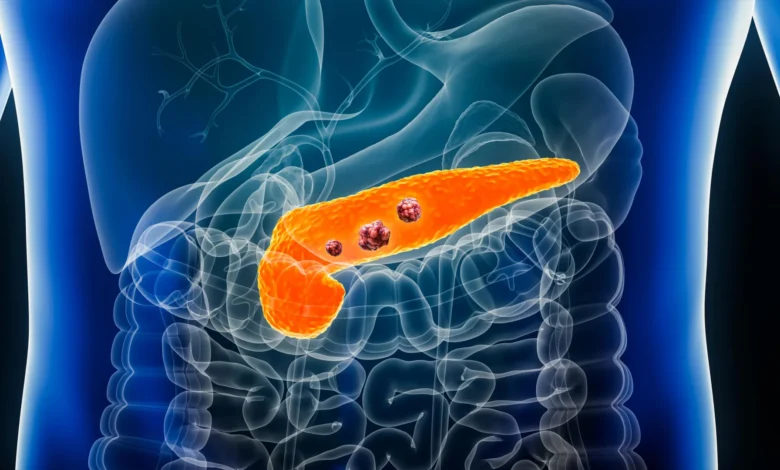
The landscape of oncology may be on the cusp of a paradigm shift, as a new artificial intelligence framework demonstrates the ability to identify the earliest, most subtle indicators of pancreatic ductal adenocarcinoma—the most lethal and common form of pancreatic cancer—years before the disease becomes visible on standard clinical imaging. Published in the journal Gut, this research introduces the Radiomics-based Early Detection MODel (REDMOD), an automated system that has outperformed human specialists in detecting physiological tissue shifts that typically elude conventional radiological review.
Pancreatic ductal adenocarcinoma is notorious for its poor prognosis, largely because it remains clinically silent during its formative stages. By the time most patients present with symptoms—such as jaundice, abdominal pain, or unexplained weight loss—the cancer has often progressed beyond the point of surgical resection. The development of REDMOD seeks to bridge this critical detection gap, potentially shifting the clinical standard from reactive treatment to proactive pre-clinical intervention.
The Challenge of Early Detection
The anatomy of the pancreas presents a significant diagnostic hurdle. Situated deep within the abdomen, the organ is often obscured by overlying bowel gas and other internal structures, making it difficult to visualize clearly on standard computed tomography (CT) scans. Furthermore, early-stage pancreatic cancer often manifests as microscopic changes in tissue texture that do not necessarily alter the organ’s gross morphology.
Radiologists are trained to look for focal masses or structural irregularities, but early-stage disease frequently leaves these features absent. Because the cancer progresses with rapid aggression, the window for successful intervention is narrow. Historically, the failure to detect these minute changes has resulted in a dismal five-year survival rate for pancreatic cancer patients, a statistic that has remained stubbornly low for decades.
REDMOD addresses this by leveraging the field of radiomics, which involves the extraction and analysis of massive amounts of quantitative data from medical images—data that are imperceptible to the human eye. By analyzing the "texture" of the pancreas at a pixel-by-pixel level, the model identifies patterns associated with malignancy that have previously been categorized as biological "noise" or normal tissue variation.
Methodology and Chronology of the Study
To test the efficacy of the REDMOD framework, researchers conducted a retrospective study utilizing abdominal CT scans from 219 patients who had been treated at multiple medical centers. The design of the study was rigorous: all patients included in the experimental group had received "normal" reports from experienced radiologists at the time their scans were taken. However, all these individuals were subsequently diagnosed with pancreatic ductal adenocarcinoma at a later date.
The timeline of the data collection spanned several years to capture the evolution of the disease:
- 87 patients (40%) were scanned 3 to 12 months prior to their eventual diagnosis.
- 76 patients (35%) were scanned 12 to 24 months before diagnosis.
- 56 patients (25%) underwent imaging up to 36 months before their diagnosis.
The researchers compared these images against a control group of 1,243 individuals who remained cancer-free for at least three years following their initial scans. To ensure the validity of the comparison, the control group was carefully matched for age, sex, and the specific dates of imaging to account for technological variations in CT equipment over time.
Performance Analysis: AI vs. The Radiologist
The results of the comparative analysis were stark. While human radiologists, despite their expertise, failed to identify early markers of disease in the majority of these pre-diagnosis scans, REDMOD demonstrated a significant diagnostic advantage.
In terms of sensitivity—the ability to correctly identify those with the disease—REDMOD achieved a 73% success rate, compared to the 39% rate achieved by the radiologists. The performance gap widened as the timeline lengthened. When analyzing scans taken more than two years prior to a clinical diagnosis, the AI maintained a 68% detection rate, while human radiologists identified signs of the disease in only 23% of cases.
Beyond sensitivity, the system demonstrated high specificity, meaning it was effective at not incorrectly flagging healthy patients. In an independent validation cohort of 539 patients, REDMOD successfully classified over 81% of scans as cancer-free. Furthermore, when applied to a public dataset from the National Institutes of Health (NIH-PCT), the model correctly identified 87.5% of healthy cases.
A key feature of REDMOD is its automation. Unlike traditional methods that require a radiologist to manually outline the pancreas to evaluate its boundaries, the model performs this task automatically. This standardization eliminates the subjective variability inherent in manual segmentation, ensuring that the diagnostic process remains consistent regardless of who is operating the software.
Clinical Implications and Future Directions
The implications of these findings for public health are profound. Current medical guidelines do not recommend routine screening for pancreatic cancer in the general population, primarily due to the high cost and low sensitivity of current imaging techniques. However, if a model like REDMOD can be integrated into routine clinical workflows—particularly for high-risk populations—it could revolutionize patient outcomes.
Researchers specifically point to the potential of targeting patients with recently diagnosed diabetes or those exhibiting unexplained weight loss. These groups are at a higher statistical risk for pancreatic malignancy. By applying AI-driven radiomics to the existing scans of these patients, clinicians could identify early-stage (Stage 0) lesions that are currently being missed.
Modeling studies suggest that if the proportion of localized pancreatic ductal carcinomas detected at an early stage could be increased from 10% to 50%, survival rates would more than double. This underscores the researchers’ assertion that the timing of diagnosis is the single most critical determinant of survival in oncology.
Limitations and the Need for Prospective Validation
Despite the optimism surrounding these results, the scientific community maintains a cautious stance. The current study was retrospective, meaning it relied on historical data. To prove its clinical utility, REDMOD must undergo prospective validation. This involves testing the system in a real-world, clinical setting where patients are screened in real-time, and the AI’s predictions are compared against actual biological outcomes as they occur.
Additionally, the study authors highlighted a demographic limitation: the patient population lacked sufficient ethnic diversity. Because genetic and environmental factors can influence the radiological appearance of pancreatic tissue, the model’s performance must be evaluated across diverse populations to ensure that it does not exhibit bias or reduced accuracy in certain ethnic groups.
Furthermore, the integration of such technology into hospitals will require significant changes in infrastructure. Radiologists and oncologists will need training to interpret AI-generated insights, and medical institutions will need to establish protocols for how to handle a "positive" result from an AI in the absence of a confirmed clinical diagnosis.
A New Era of Proactive Interception
The success of the REDMOD framework represents a broader movement in medicine: the shift from "symptomatic diagnosis" to "proactive pre-clinical interception." By treating the human body as a source of high-dimensional data that can be interpreted through machine learning, researchers are beginning to uncover patterns of disease that were previously invisible.
While REDMOD is not yet ready for daily clinical use, the study provides a "proof of concept" that is difficult to ignore. If future validation confirms that the model can reliably identify stage 0 cancer, it would provide a much-needed tool in the fight against one of the most aggressive diseases in medicine.
The researchers conclude that the framework offers "tangible hope" for a field that has seen little progress in early detection. As computational power continues to grow and AI models are trained on larger, more diverse datasets, the ability to "see" into the future of a patient’s health—and intervene before a diagnosis becomes a terminal reality—is moving from the realm of science fiction into the reach of clinical possibility. The next phase of research, focusing on prospective trials and broader demographic testing, will be the ultimate test of whether this technological leap can be translated into increased survival rates for patients worldwide.
Related posts: