
The Evolution of Jessi Draper From Hairstylist to Global Influencer and the Strategic Expansion of the MomTok Brand
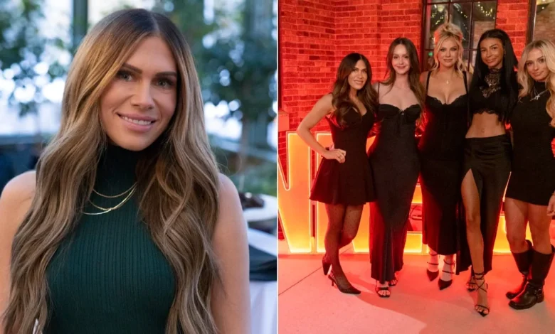
Jessi Draper, a prominent figure in the viral "MomTok" community and a breakout star of the Hulu original series The Secret Lives of Mormon Wives, has formalized a strategic partnership with the beauty brand Raw Sugar, marking a significant milestone in her transition from a regional hairstylist to a national influencer and business mogul. Amidst the ongoing production flux and high-profile speculation surrounding the future of the Mormon Wives franchise, Draper has leveraged her platform to advocate for accessible hair care and advanced dermatological routines. This development comes as the reality television landscape increasingly serves as a primary incubator for consumer brand growth, particularly in the beauty and wellness sectors. Draper, who has long maintained a professional career in the hair industry within Utah, is now utilizing her expertise to bridge the gap between salon-grade results and affordable retail products, emphasizing a holistic approach to scalp health and skincare.
The Cultural and Media Context of the MomTok Phenomenon
To understand the significance of Draper’s recent business ventures, it is essential to examine the trajectory of the "MomTok" collective. The group initially garnered international attention in 2022 following a series of internal scandals and revelations regarding "soft swinging" within a subset of the community in Utah. What began as a localized social media niche quickly transformed into a cultural touchstone, prompting Hulu to greenlight a docuseries titled The Secret Lives of Mormon Wives. The series, which premiered in September 2024, explores the complex intersection of traditional religious values, modern motherhood, and the high-stakes world of social media influence.
Draper has emerged as a stabilizing force within the cast, balancing the inherent drama of reality television with a clear focus on her professional credentials. As a veteran hairstylist, her insights into the beauty industry carry a level of authority that resonates with a consumer base increasingly skeptical of celebrity endorsements lacking technical background. The success of the series has created a robust economic ecosystem for its participants, with Draper leading the charge in securing partnerships that align with her established career path.
Strategic Brand Alignment: The Raw Sugar Partnership
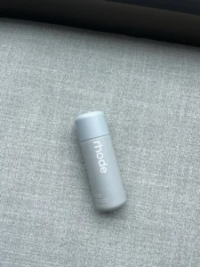
The collaboration between Jessi Draper and Raw Sugar represents a calculated move toward "affordable luxury" in the personal care market. Raw Sugar, a brand known for its "Cold-Press Technology" and commitment to clean ingredients, has historically targeted the mass-market demographic through major retailers such as Target and Amazon. By partnering with an expert hairstylist like Draper, the brand gains professional validation for its formulations.
:quality(85):upscale()/2026/04/16/773/n/1922153/a97c122869e11d89c4f016.09521510_.png)
Draper has specifically highlighted the brand’s Scalp Restore line, which includes a shampoo and conditioner priced at approximately $11.00 each. From a journalistic and industry perspective, this partnership taps into the "skinification of hair" trend—a market shift where consumers treat their scalp with the same level of sophistication and care previously reserved for facial skincare. Draper noted that as a professional, her primary concern is the prevention of follicular clogging and chemical buildup, which can be mitigated through regular scalp detoxing and the use of pH-balanced products.
Industry data suggests that the scalp care market is projected to grow significantly over the next five years. By focusing on scalp health rather than just aesthetic styling, Draper is positioning herself at the forefront of a science-backed beauty movement. She emphasizes that scalp treatments, ideally performed once or twice a year, are critical for maintaining hair integrity, especially for those who frequently use styling products or live in climates that contribute to dryness.
Chronology of Jessi Draper’s Rise and Brand Development
The timeline of Draper’s professional evolution reflects the broader shifts in the digital economy:
- Pre-2022: Jessi Draper establishes herself as a high-end hairstylist and business owner in Utah, specializing in extensions and color.
- May 2022: The "MomTok" community faces intense public scrutiny following viral disclosures of internal group dynamics. Draper remains a core member of the social circle while maintaining her professional practice.
- Early 2023: Production begins on The Secret Lives of Mormon Wives. Draper begins to integrate her professional hair care advice with her growing social media presence.
- September 2024: The Hulu series launches to high viewership numbers, making the cast household names within the reality TV genre.
- Late 2024: Draper moves into a new residence in Utah and announces her partnership with Raw Sugar, signaling a shift toward national brand ambassadorship.
- Present: Draper continues to expand her influence, incorporating K-Beauty and high-end cosmetics into her public-facing beauty regimen while awaiting news on the series’ second season.
Advanced Skincare and the Influence of K-Beauty
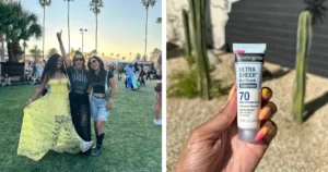
In addition to her hair care advocacy, Draper has become a vocal proponent of the South Korean beauty (K-Beauty) industry, specifically the brand Medicube. This shift reflects a broader consumer trend toward "glass skin" aesthetics and high-tech home skincare devices. Draper has credited Medicube’s product line, particularly the PDRN Pink Caffeine Night Wrapping Mask, with transforming her skin texture.
The inclusion of PDRN (Polydeoxyribonucleotide), an ingredient often derived from salmon DNA known for its regenerative properties, highlights Draper’s interest in advanced dermatological ingredients. Market analysis indicates that K-Beauty continues to dominate the global skincare conversation due to its innovative use of ingredients and focus on long-term skin health. Draper’s endorsement of these products, alongside her use of Charlotte Tilbury’s "Kissing Satin Shine" lipsticks and GrandeLash growth serums, showcases a curated blend of high-street affordability and luxury performance.
:quality(70):extract_cover():upscale():fill(ffffff)/2026/04/16/774/n/1922153/ddfee35c69e11db8949a15.20146698_Screenshot_2.png)
The Business of Reality TV: Makeup and On-Screen Presentation
A significant aspect of Draper’s evolution involves the technical requirements of being a television personality. She has noted that the transition from social media filters to high-definition television cameras required a complete overhaul of her makeup application techniques. Colors and textures translate differently under professional studio lighting compared to natural light or mobile phone cameras.
Draper has admitted to a growing "addiction" to professional makeup artistry, a common sentiment among reality stars who must maintain a "camera-ready" appearance for grueling filming schedules. This has led to an increased interest among her followers in the specific "hacks" used by the cast. For instance, Draper adopted a specific eyelash routine from fellow cast member Miranda McWhorter, which includes the use of lash-enhancing serums to achieve a look of fullness that survives the demands of the screen.
Extension Technology and the Utah Aesthetic
As an expert in hair extensions, Draper has also addressed the "Utah aesthetic," which often features long, voluminous hair. She posits that extensions are no longer merely about length but are a functional tool for achieving "fullness" and ensuring that styles hold their shape throughout the day. This perspective reframes extensions as a practical hair accessory rather than a purely cosmetic vanity item. She recommends the consistent use of hair serums to moisturize the mid-lengths and ends of extensions, particularly in the arid climate of the Mountain West, which can be devastating to hair moisture levels.
Psychological Impact and the "Respond, Don’t React" Philosophy
Beyond beauty and business, Draper has spoken candidly about the psychological toll of reality television fame. The genre often rewards explosive emotional reactions, yet Draper has publicly committed to a philosophy of "responding rather than reacting." This approach is a strategic move to preserve her professional reputation and mental well-being in an environment designed for conflict.
She has integrated rigorous self-care routines into her daily life to combat the stress of public scrutiny. This includes a nightly ritual involving Epsom salt baths, aromatherapy, and "unplugging" from social media. This emphasis on mental health is a recurring theme among modern influencers who face the "always-on" pressure of the digital economy. Draper’s advice to her younger self—to view mistakes as growth opportunities—reflects a maturing brand identity that prioritizes longevity over short-term viral moments.
:quality(70):extract_cover():upscale():fill(ffffff)/2026/04/16/774/n/1922153/0edc8e0869e11ddc54a007.38872399_Screenshot_2.png)
Broader Economic and Social Implications
The trajectory of Jessi Draper and the MomTok collective offers a case study in the power of "niche-to-mainstream" pipeline dynamics. The Mormon influencer market is a multi-million dollar industry, characterized by high engagement rates and a demographic that is highly coveted by advertisers. By maintaining a foot in both the traditional professional world (hair styling) and the new media world (Hulu/TikTok), Draper provides a blueprint for how reality stars can build sustainable careers that outlast their time on screen.
The partnership with Raw Sugar is likely the first of many such deals as the cast of The Secret Lives of Mormon Wives continues to navigate their newfound fame. As the "Mormon Wives universe" evolves, the focus will likely shift from the scandals that launched the group to the individual brands they build. Draper’s focus on scalp health, K-Beauty, and professional-grade hair care suggests a brand built on expertise and reliability—a potent combination in the current influencer market.
While the future of the Hulu series remains "up in the air" in terms of official renewal announcements, the individual cast members are not waiting for a production green light to advance their commercial interests. Draper’s "booked and busy" status is a testament to the fact that in the modern media landscape, the platform is the starting point, but the individual’s professional utility is what determines their ultimate success. As she continues to move between her home state of Utah and the national stage, Jessi Draper remains a central figure in the ongoing dialogue regarding beauty, faith, and the business of being seen.


