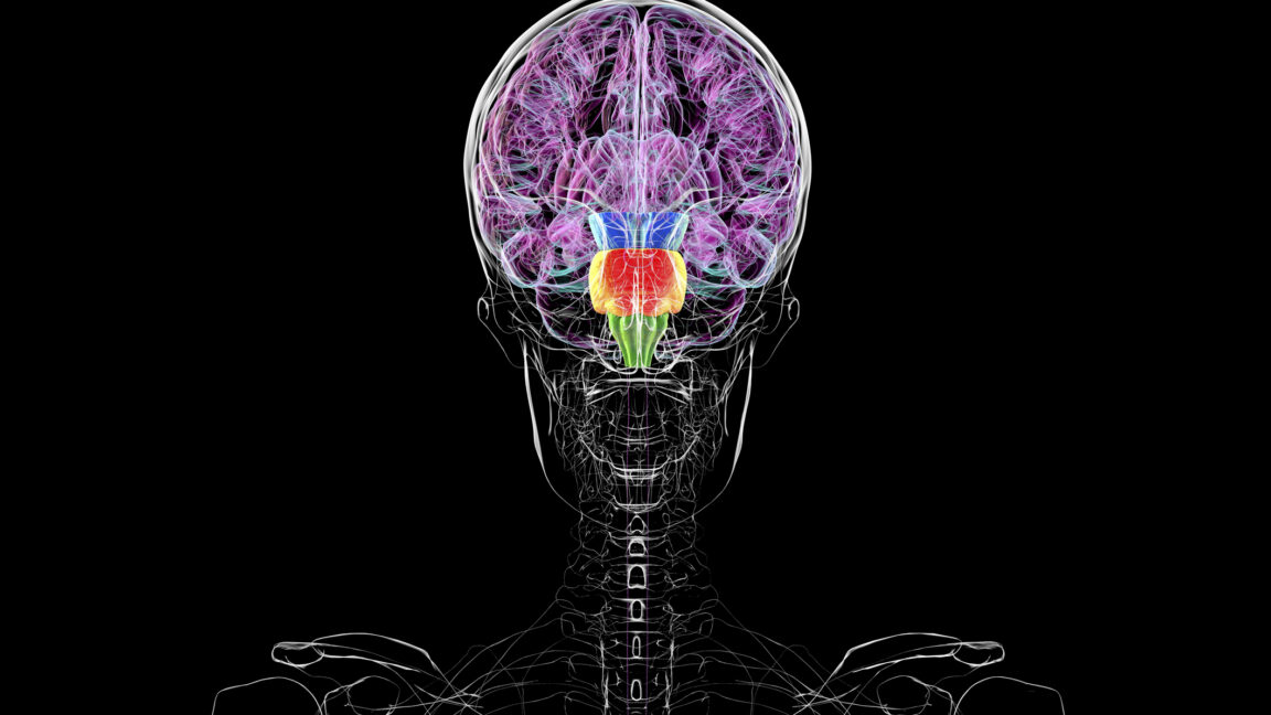
Recent headlines across mainstream media outlets have suggested a radical shift in our understanding of human anatomy, with claims that humans possess "two separate brains" rather than one unified central nervous system. These reports, which originated from a single university press release disseminated late last week, have sparked confusion regarding neurobiology. To clarify the record: human beings do not possess two brains. However, the underlying research study provides a sophisticated, high-resolution look at how the vertebrate nervous system segments itself during early embryonic development. By deconstructing the mechanisms that differentiate the forebrain and midbrain from the hindbrain, this research offers a compelling case study in developmental biology, illustrating how a single sheet of undifferentiated cells matures into the most complex organ in the body.
The Origins of a Misunderstanding
The scientific curiosity surrounding this topic stems from a fundamental question in embryology: how does the nervous system organize itself into distinct functional regions? The recent research, which utilizes advanced fluorescent labeling in murine (mouse) models and human pluripotent stem cells, identifies a critical, early-stage genetic divide in the developing ectoderm.
The media frenzy began when a university press release described the distinct genetic signatures of the hindbrain versus the forebrain and midbrain as "two brains." In reality, the study highlights that as early as the neural plate stage, cells are already "fated" to become specific regions of the central nervous system. While the brain is physically continuous, its developmental history is compartmentalized. This is not a discovery of a second brain, but rather a discovery of how early "positional information" dictates the architectural blueprint of the developing skull and spinal cord.
Chronology of Development: From Ectoderm to Neural Tube
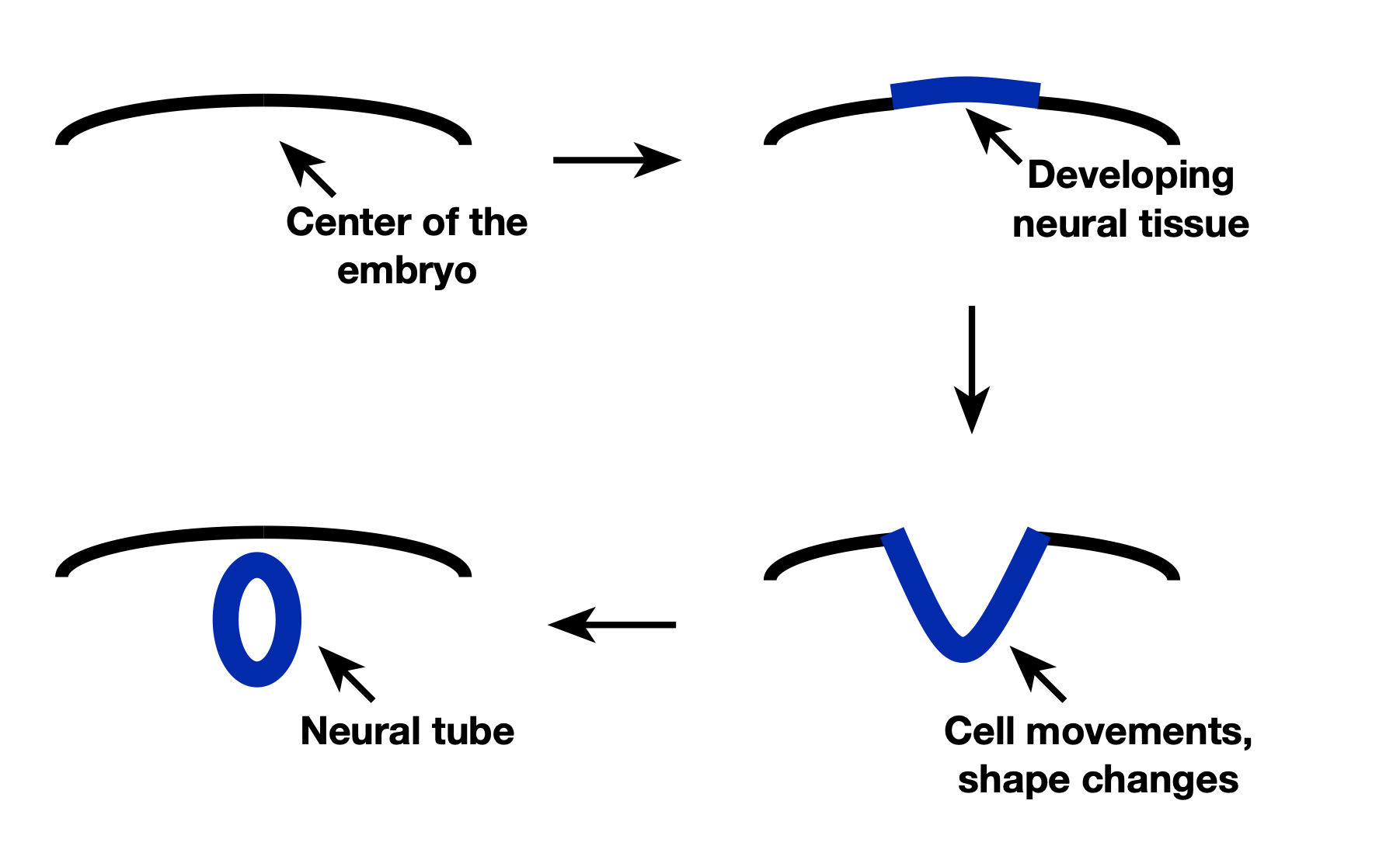
To understand the significance of this research, one must first look at the timeline of vertebrate development. Roughly three weeks after fertilization in humans, the process of neurulation begins. At this stage, the embryo is composed of three primary germ layers: the endoderm, the mesoderm, and the ectoderm.
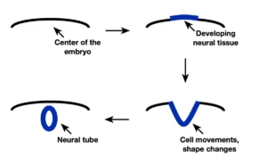
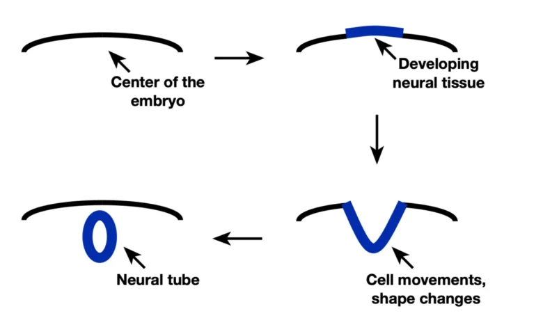
The ectoderm is the progenitor of the entire nervous system. Through a process called primary neurulation, a specialized strip of ectoderm cells—the neural plate—thickens and begins to fold. Over several days, the edges of this plate rise and meet in the midline, fusing to form the neural tube. This tube is the precursor to the brain and spinal cord.
Crucially, this study demonstrates that even before the neural tube is fully formed, the cells within the neural plate have already received and processed molecular signals that distinguish them as either "future hindbrain/spinal cord" or "future forebrain/midbrain." This "fate mapping" ensures that when the tube closes, the regional identity of the cells is already locked in, allowing for the precise, sequential development of specialized neural structures.
Data and Methodology: Fluorescent Fate Mapping
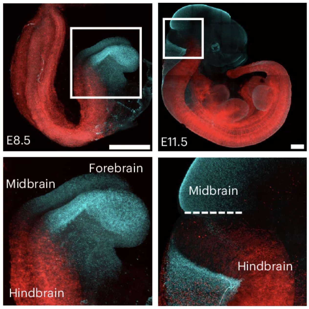
The researchers employed a technique known as "lineage tracing" to visualize these early divisions. By modifying specific genes—specifically those responsible for early spatial patterning—they enabled cells in the anterior (front) half of the embryo to express a cyan fluorescent protein, while cells in the posterior (back) half expressed a red fluorescent protein.
The data revealed a striking degree of consistency:
- Segmental Fidelity: In 96 percent of the observed cases, the descendants of individual progenitor cells remained within their assigned segment (either the hindbrain/spinal cord or the midbrain/forebrain).
- Human Validation: The researchers corroborated these findings using human-derived stem cells, which were exposed to morphogens—signaling molecules that govern the spatial arrangement of tissues. The results showed that human stem cells follow a similar, albeit more complex, developmental logic to that of mice.
- The Border Effect: The four percent of cases where cells crossed the boundary suggest a level of "plasticity" at the interface, where cells might be influenced by neighboring environments if they migrate early enough.
The Role of Morphogens and Positional Identity
The implications of this early-stage "segmentation" are profound. By establishing regional identity early, the nervous system becomes much more efficient at utilizing signaling molecules. One of the most famous of these is the Sonic Hedgehog (SHH) protein.
If all neural cells were identical, a single signal like SHH might trigger the same response across the entire brain. However, because the hindbrain and forebrain have inherited different "developmental histories," they interpret these signals differently. This is known as "context-dependent signaling." A cell in the midbrain receiving a BMP (Bone Morphogenetic Protein) signal will activate a different set of genes than a cell in the spinal cord receiving that exact same signal.
This mechanism allows the embryo to generate immense complexity with a relatively small toolkit of signaling molecules. By recycling these signals at different stages—first to determine fate, then to guide migration, and finally to facilitate synaptic wiring—the body avoids the need for a separate, unique signaling molecule for every single biological process.
Scientific Consensus and Peer Response
While the "two brains" narrative has been dismissed by the broader scientific community, the underlying research has been lauded for its methodological rigor. Experts in developmental biology note that this study addresses a long-standing "black box" in the field: the transition from a homogenous-looking sheet of cells to a highly heterogeneous central nervous system.
"The study does not show we have two brains," noted one independent embryologist familiar with the work. "It shows that the brain is not a uniform piece of tissue that gets divided up later. It is a mosaic that begins its existence as a pre-programmed collection of distinct segments. This is a vital contribution to our understanding of birth defects and neurodevelopmental disorders, where the failure of these boundaries to form correctly can lead to significant structural anomalies."
Broader Implications for Medicine and Research
The ability to map these boundaries has significant implications for regenerative medicine. As scientists continue to develop organoids—lab-grown, three-dimensional tissues that mimic brain development—understanding these early "fate" cues is essential. To grow a functional midbrain-like organoid, researchers must first understand the precise timing and sequence of signals that the embryo uses to differentiate that specific region from the hindbrain.
Furthermore, this research provides a window into the evolution of vertebrates. By comparing these markers across species, biologists can trace how different animals have adapted the basic vertebrate "tube" plan to create diverse neurological capabilities, from the relatively simple nervous system of a tadpole to the highly folded, complex cortex of a human.

Conclusion: One Organ, Many Histories
The human brain remains a single, unified command center, but it is an organ built through a series of distinct, precisely choreographed steps. The recent study published by the researchers at Stanford does not challenge the unity of the human brain; rather, it elevates our appreciation for the complexity of its construction.
By identifying the early molecular markers that separate the hindbrain from the midbrain, the study confirms that our neurological architecture is established long before the first thought is ever formed. The "two brains" headline, while factually incorrect, serves as an accidental entry point into one of the most fascinating aspects of biological science: the transformation of a simple, flat layer of cells into the sophisticated, segmented, and highly efficient engine of human consciousness. As research continues to refine our understanding of these developmental boundaries, the focus will remain on how these early genetic "maps" translate into the complex wiring that allows us to perceive, act, and think.
Related posts: