Silent Memory Thieves: How Tau Protein and Disrupted Brain Waves During Deep Sleep Compromise Cognitive Health

The experience of wandering through a crowded parking lot, unable to recall where one parked, is a universal frustration often dismissed as a byproduct of aging. However, recent groundbreaking research from the University of California, Berkeley, suggests that these minor lapses may be the tip of an iceberg linked to the subtle, progressive biological changes associated with Alzheimer’s disease. Published in the journal Nature Neuroscience, the study reveals that the accumulation of the protein tau—a hallmark of Alzheimer’s pathology—directly interferes with the brain’s ability to coordinate electrical activity during deep, non-REM sleep, effectively fracturing the mechanisms that consolidate and preserve our memories.
The Anatomy of Memory Consolidation
Episodic memory, the cognitive system responsible for recording the "what, where, and when" of our daily experiences, is notoriously fragile. From the vivid recollection of a childhood milestone to the mundane task of remembering a parking spot, these memories must be "consolidated" overnight to transition from short-term storage to long-term retrieval. This process occurs predominantly during deep sleep, a stage characterized by slow, rhythmic electrical oscillations.
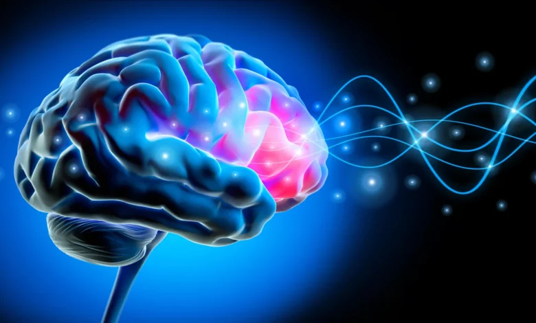
During these hours, large populations of neurons in the frontal cortex fire in a synchronized, cascading sequence. These "slow waves" act as a biological orchestra, ensuring that information gathered during the day is processed and stabilized. However, as the human brain ages, this internal rhythm often falters. For years, the scientific community has grappled with why memory performance declines in older adults, even in those who remain cognitively healthy. The UC Berkeley team, led by neuroscientist Omer Sharon and former sleep researcher Matthew Walker, has provided a critical piece of the puzzle: the physical obstruction of these waves by tau protein.
The Phenomenon of Lonely Waves
The researchers utilized electroencephalograms (EEGs) to monitor the sleeping brain, comparing a cohort of healthy young adults in their early 20s with an older group aged 65 to 75. The findings were stark. In younger brains, slow waves traveled across the scalp in broad, sweeping, and highly coordinated patterns, covering distances roughly equivalent to the width of a human hand.
In contrast, the older participants exhibited what researchers have termed "lonely waves." Instead of migrating across the brain in cohesive, long-range bursts, these electrical pulses appeared isolated, traveling over much shorter distances and failing to reach the necessary synchronization to effectively "write" memories into the brain’s long-term storage. The result is a failure of overnight memory consolidation, which directly correlates to poorer performance in recall tests conducted the following day.
Tau Protein and the Pathological Link
To determine if this decline was linked to Alzheimer’s-related pathology, the team collaborated with Dr. William Jagust, an expert in public health and neuroscience. Using positron emission tomography (PET) scans, the researchers mapped the distribution of tau protein in the brains of the older participants. The correlation was undeniable: higher concentrations of tau in the frontal cortex—the origin point for these global sleep waves—predicted the fragmentation of the waves.
Crucially, this study highlights that these pathological changes often precede the clinical diagnosis of Alzheimer’s disease. "These people did not have Alzheimer’s," explained Omer Sharon. "They had tau in their brains, but with subclinical impacts. Their memory decline was within the normal range for their age." This indicates that the mechanisms of cognitive decline are set in motion long before the onset of symptomatic dementia, suggesting that the "normal" decline in memory as we age may be a spectrum of pathology rather than an inevitable, uniform process.
Multi-Modal Evidence and Validation
To bolster the validity of their findings, the Berkeley team sought to confirm the relationship between protein accumulation and sleep disruption through an alternative biological marker. By collaborating with neurologist Yo-El Ju at Washington University in St. Louis, the researchers analyzed spinal fluid from an independent cohort of older adults.
While PET scans provide a localized view of tau in the brain, spinal fluid analysis offers a systemic look at the ratio of tau to amyloid-beta, another protein synonymous with Alzheimer’s development. The results mirrored the imaging data: participants who showed higher ratios of these proteins in their spinal fluid were statistically more likely to exhibit the "lonely wave" pattern during sleep. This cross-methodological consistency provides a robust foundation for the theory that neurodegenerative markers are physically impeding the brain’s recovery and memory-storage systems.
A Longitudinal Perspective
The research did not merely capture a static snapshot of the brain; the team tracked a subset of participants over several years to observe the trajectory of their cognitive health. This longitudinal analysis revealed a clear, worsening correlation: as tau levels in the frontal cortex rose over time, the slow waves became increasingly disorganized, and the subjects’ capacity to retain newly learned information decreased proportionately.
This data provides a compelling narrative for the progression of memory loss. While it does not definitively prove that tau is the primary "cause" of the wave disruption, it establishes a functional link that characterizes the early stages of cognitive impairment. The question remains as to whether the breakdown of these waves exacerbates the buildup of tau, or if the protein accumulation is the sole initiator. Disentangling this "chicken-or-egg" scenario is now the primary focus for future neurological research and potential therapeutic intervention.
Broader Implications for Clinical Neurology
The implications of this research are profound for the field of neurology and the development of future dementia treatments. If deep-sleep disruption is a primary mechanism through which tau protein erodes memory, then identifying and treating sleep disturbances could become a frontline defense against cognitive decline.
Currently, diagnostic tools for Alzheimer’s are often invasive or prohibitively expensive, relying on PET scans or lumbar punctures. If clinicians could reliably use high-density EEG monitoring to identify "lonely waves" as a proxy for early tau accumulation, it could revolutionize screening processes. Early detection could allow for the administration of emerging anti-tau or anti-amyloid therapies at a stage where the brain still retains significant plasticity.
Furthermore, the study challenges the notion that memory loss is an unavoidable consequence of chronological aging. Instead, it frames memory health as a manageable biological process dependent on the integrity of the brain’s nighttime "housekeeping" systems. By highlighting the role of the frontal cortex as the command center for these sleep waves, researchers have identified a specific target for both diagnostic and potentially restorative interventions.
Future Research Directions
The scientific community is now poised to move from observation to intervention. The next phase of research will likely focus on whether external stimuli—such as neurostimulation techniques that can boost or synchronize slow waves—could mitigate the impact of tau in the aging brain. If the "orchestra" of the brain can be artificially re-synchronized, can the memory deficits be slowed or even reversed?
The collaborative effort, which included contributions from researchers such as Xi Chen, James Westphal, and Chelsea Brown, has set a new standard for how we view the relationship between sleep architecture and neurodegeneration. With funding from the National Institute on Aging at the National Institutes of Health, as well as support from the Glenn Foundation for Medical Research and the Weill Neurohub, the team has successfully shifted the conversation from general "sleep quality" to the specific, measurable electrical failures that define the onset of cognitive disease.
As the global population ages, understanding the biological basis of memory loss is no longer just a matter of academic interest; it is a public health imperative. The discovery of the link between tau-induced pathology and the disruption of synchronized brain waves provides a roadmap for future research, offering a flicker of hope that, with enough scientific clarity, the "lonely waves" of the aging brain might one day be brought back into harmony.