Groundbreaking Discovery of Universal Stem Cells Offers New Hope for Treating Spinal Stenosis and Connective Tissue Disorders

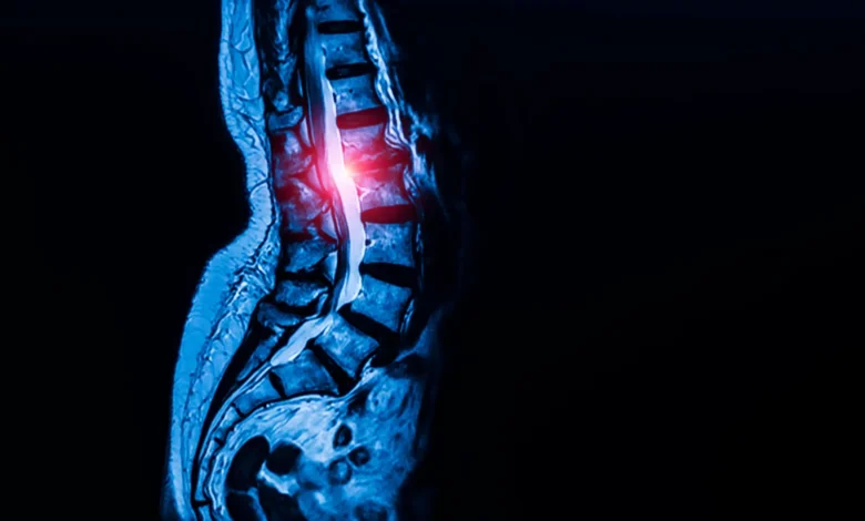
A team of researchers at Weill Cornell Medicine and the Hospital for Special Surgery (HSS) has successfully identified a previously elusive population of stem cells responsible for the formation and maintenance of the body’s tendons and ligaments. This discovery, published in the September 7 edition of the journal Cell, marks a significant milestone in musculoskeletal biology, providing a potential biological roadmap for addressing conditions that have long been managed primarily through invasive surgical intervention. By isolating the progenitor cells that build these connective tissues, the scientific community now has a targeted pathway to investigate, and potentially reverse, the debilitating effects of lumbar spinal stenosis, a condition that impacts approximately 103 million people globally.
The identification of these cells represents the culmination of a multi-year effort to map the skeletal system’s cellular architecture. While previous research had successfully identified stem cells associated with bone fracture repair, skull formation, and general spinal development, the specific stem cell population governing tendons and ligaments had remained elusive. The difficulty stemmed from the complex, fibroblast-rich environment of these tissues, where specialized cells often share similar morphological characteristics with their mature counterparts, making isolation exceptionally challenging.
The Methodology of Discovery: Mapping the Cellular Landscape
The research team, led by co-corresponding authors Dr. Matthew Greenblatt, an associate professor of pathology and laboratory medicine at Weill Cornell Medicine, and Dr. Sravisht Iyer, a spine surgeon at HSS, utilized advanced single-cell analysis to differentiate these specific stem cells from the surrounding cellular matrix. The process involved analyzing thousands of individual cells and sorting them based on their functional properties, specifically their "stemness"—the capacity for both self-renewal and the generation of the full spectrum of tendon and ligament cell types.
Initial studies were conducted in murine models, where researchers identified these cells residing in specialized niches within tendons and ligaments. These niches serve as a reservoir for tissue growth and repair. Upon validating the existence of these cells in mice, the team moved to human samples. Dr. Lingling Hu, a postdoctoral fellow and the study’s first author, spearheaded the effort to isolate these cells from human ligament tissue harvested during consented surgeries. The team confirmed that these human-derived cells possessed the same dual capacity for self-renewal and differentiation observed in the animal models.
Beyond the spine, the researchers conducted a comprehensive survey of the body’s connective tissues. By examining samples from the patellar (kneecap) ligament and the Achilles tendon, the team concluded that this stem cell population appears to be universal, functioning as the fundamental building block for tendons and ligaments throughout the entire human musculoskeletal system.
The Pathological Link: Spinal Stenosis and Calcium Signaling
Lumbar spinal stenosis occurs when the spinal canal narrows, causing pressure on the nerves. This constriction is frequently the result of the thickening of spinal ligaments. As these ligaments expand, they reduce the available space within the spinal canal, leading to chronic pain, numbness, and severe mobility impairment. For many patients, the current clinical standard is to wait for the condition to progress to a point where surgical decompression is the only viable option.
The research team sought to determine if the newfound stem cells were contributing to this pathological thickening. By comparing stem cells derived from patients suffering from spinal stenosis against those from patients with non-stenotic herniated discs, the investigators discovered a marked disparity. Ligaments taken from patients with stenosis contained a significantly higher concentration of these stem cells. Furthermore, when these "stenosis-associated" cells were transplanted into mouse models, they exhibited a propensity for excessive tissue production compared to healthy control cells.
A closer analysis of the intracellular mechanisms revealed that these hyperactive stem cells were governed by elevated calcium signaling. Calcium signaling is a critical regulatory pathway that controls cellular growth, metabolism, and communication. The researchers discovered that by genetically modulating this signaling pathway, they could influence the growth rate of the tissue. When they artificially increased calcium signaling in healthy cells, the cells began to produce excessive, pathological tissue growth. Conversely, by inhibiting this signaling in a mouse model of spinal stenosis, the researchers successfully blocked the abnormal growth process.
Clinical Implications and Potential Therapeutic Targets
The revelation that calcium signaling acts as a "master switch" for these stem cells opens a new frontier in pharmacology. Dr. Iyer noted that this is among the first studies to identify a clear, actionable therapeutic target for a condition that has historically been treated with a "wait and watch" approach.
One of the most promising aspects of this discovery is the potential repurposing of existing medications. Calcium channel blockers, a class of drugs currently used to manage hypertension, are well-understood, have established safety profiles, and are widely available. While the research team emphasizes that clinical trials are essential to determine whether these drugs can effectively halt or reverse spinal stenosis in humans, the biological evidence provides a strong rationale for immediate further study.
The shift from reactive surgery to proactive, mechanistic treatment could fundamentally transform spinal care. By identifying the underlying cellular dysfunction before severe nerve compression occurs, clinicians may be able to preserve patient mobility and reduce the need for high-risk surgical procedures.
Broader Impact and Future Research Directions
The implications of this study extend far beyond spinal health. Given that these cells are the origin point for all tendons and ligaments, the researchers anticipate that defects in this cell population are likely at the core of a wide array of connective tissue disorders. Future research initiatives are already being planned to investigate the role of these stem cells in conditions such as:
- Marfan Syndrome: A genetic disorder affecting connective tissues throughout the body, including the heart, blood vessels, and skeletal system.
- Rotator Cuff Injuries: Exploring why these common shoulder injuries are frequently resistant to standard healing protocols.
- Achilles Tendinopathy: Examining chronic degeneration and the failure of natural repair mechanisms.
- Ligament Reconstruction: Developing biological scaffolds or targeted therapies to improve outcomes in ACL and other major ligament repair surgeries.
Scientific Context and Funding
The study was supported by a wide array of prestigious organizations, reflecting the significance of the findings in the fields of orthopedics and regenerative medicine. Funding partners included the Marfan Foundation, the National Institutes of Health (NIH), the Arthritis National Research Foundation, and the Pershing Square Foundation, among others.
This research arrives at a time when the global burden of musculoskeletal disease is increasing due to aging populations. As researchers move toward the next phase—human clinical trials and large-scale longitudinal studies—the medical community remains cautiously optimistic. By moving the focus from the mechanical symptom (narrowing of the canal) to the cellular driver (stem cell hyperactivity), the Weill Cornell and HSS team has provided a blueprint that could define the next generation of orthopedic intervention.
While the road to a clinical application involves rigorous testing, the identification of this universal stem cell provides a level of clarity that has been missing for decades. The ability to manipulate these cells, either to suppress pathological growth in stenosis or potentially to stimulate repair in degenerative injuries, represents a fundamental shift in our understanding of how the body maintains its structural integrity. As Dr. Greenblatt and Dr. Iyer continue their work, the focus will remain on translating these molecular findings into concrete, life-altering treatments for millions of patients worldwide.