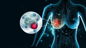
The Surprising Role of Dietary Fructose in Aggressive Ovarian Cancer Metastasis and Potential Implications for Treatment

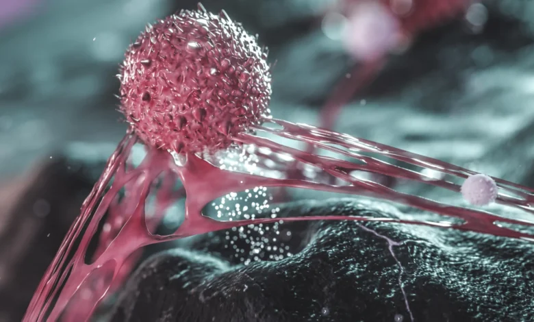
A groundbreaking study conducted by researchers at The Wistar Institute has uncovered a sophisticated and previously unknown mechanism by which aggressive ovarian cancer cells manipulate their environment to survive and spread throughout the body. The research, published in the journal Nature Aging, identifies a common dietary sugar—fructose—as a critical chemical signal that enables cancer cells to break free from their primary tumor site, a process that represents the most lethal phase of the disease. This discovery not only challenges our understanding of tumor microenvironments but also introduces complex questions regarding the intersection of nutrition, metabolic health, and standard cancer therapies.
The Challenge of Ovarian Cancer Recurrence
Ovarian cancer remains one of the most formidable challenges in modern oncology, primarily due to its propensity for late-stage diagnosis and high rates of recurrence. Standard clinical practice involves the administration of platinum-based chemotherapy agents, such as cisplatin or carboplatin. These drugs function by inducing DNA damage within rapidly dividing cancer cells, effectively triggering programmed cell death or apoptosis.
While a significant majority of patients show an initial positive response to these regimens, the clinical reality is that the cancer frequently returns. In approximately 90% of cases, patient mortality is linked to metastasis—the process by which malignant cells migrate from the ovaries to populate the abdominal cavity, eventually invading vital organs such as the liver, intestines, and stomach. For decades, the scientific community has grappled with the question of why these cells, despite being chemically targeted and seemingly incapacitated, possess the resilience to eventually reform into aggressive, treatment-resistant tumors.
Decoding the Persistence of Senescent Cells
The Wistar Institute research team, led by Dr. Katherine Aird and postdoctoral fellow Dr. Aidan Cole, focused their inquiry on a specific subpopulation of cancer cells that survive chemotherapy. Although these cells cease to multiply—a state often referred to as senescence—they remain biologically active.
"Some cancer cells that survive chemotherapy aren’t dividing anymore, but they’re still biologically active," explained Dr. Aidan Cole. "Instead, they continue to release molecules that send signals to nearby cells. Our study is among the first to show that a nutrient—in this case, fructose—can act as one of those signals."
The research team employed advanced preclinical models to demonstrate that it is the secretome—the collection of molecules secreted by these surviving cells—rather than the cells themselves, that acts as the primary driver of metastasis. By separating the surviving cells from their released molecules in an experimental setting, the team observed that the substances alone were sufficient to induce significant spreading behaviors in nearby healthy or dormant cancer cells.
Fructose as a Molecular Messenger
The most startling aspect of the study is the identification of fructose as the signaling molecule. Unlike glucose, which is the body’s primary energy substrate, fructose—found naturally in fruits and honey, but consumed in massive quantities via high-fructose corn syrup—serves here as a strategic communication tool.
The study indicates that chemotherapy-treated cells begin to secrete higher levels of fructose into the extracellular space. This fructose does not serve as a fuel source for the neighboring cells; rather, it acts as a ligand or chemical messenger. When neighboring cancer cells detect this fructose, it triggers a metabolic cascade that effectively dismantles their ability to maintain cellular adhesion.
The Mechanism of Cellular Escape
To map the biological pathways involved, the researchers utilized CRISPR-Cas9 genome-editing technology to conduct a systematic screen. This allowed them to pinpoint the specific genes and proteins regulated by the presence of fructose. Their findings revealed that fructose actively suppresses the synthesis of cholesterol in neighboring cells.
Cholesterol is a vital building block for the lipid bilayer of cell membranes. Beyond its structural role, cholesterol is essential for the formation and maintenance of "cell-cell junctions." These junctions act as a biological glue, tethering individual cells together within the cohesive structure of a tumor. When fructose downregulates cholesterol production, these structural bonds weaken. Deprived of the necessary lipid components to maintain these tight connections, the cancer cells lose their structural integrity and detach from the primary mass, becoming mobile and capable of infiltrating surrounding abdominal tissues.
The Statin Connection: A Medical Paradox
The discovery that cholesterol suppression facilitates tumor spread immediately prompted the research team to investigate the impact of statins. Statins are among the most widely prescribed medications in the United States, used by an estimated 39 million Americans to manage cardiovascular risk by inhibiting the HMG-CoA reductase enzyme, which is central to cholesterol production.
The team’s preliminary experiments suggested that statins, when administered in a laboratory setting, could mirror the effects of fructose by weakening cell-to-cell adhesion. This finding introduces a critical area of concern for future clinical investigation: whether the use of cholesterol-lowering drugs in patients undergoing chemotherapy for ovarian cancer might inadvertently create a more favorable environment for metastasis.
Dr. Katherine Aird emphasized the urgency of these findings, particularly for the demographic most affected by the disease. "We haven’t tested this effect in patients yet, but it raises questions about combining cholesterol-lowering drugs with chemotherapy, especially since ovarian cancer is most common in postmenopausal women who are often already on statins," she noted.
Implications for Diet and Clinical Practice
While the findings provide a compelling narrative regarding the link between sugar consumption and tumor behavior, the researchers caution against drawing premature conclusions. The study establishes a mechanism in preclinical models, but it does not yet prove that dietary sugar intake directly causes ovarian cancer progression in humans.
Nonetheless, the data is striking. In the United States, high-fructose corn syrup accounts for a substantial portion of daily caloric intake, ranging between 8% and 20% for many individuals. If the relationship between fructose, cholesterol synthesis, and metastasis holds true in humans, it could pave the way for novel nutritional interventions. Future clinical trials may explore whether dietary modifications or targeted metabolic inhibitors could be used in conjunction with standard chemotherapy to "lock" cancer cells in place, preventing them from escaping and spreading.
Looking Toward a Broader Scope
The Wistar Institute team suspects that this mechanism may not be exclusive to ovarian cancer. Cancers that develop within the abdominal cavity, including pancreatic, colon, and liver cancers, share similar microenvironmental conditions. The team has already initiated follow-up studies to determine if these cancers utilize fructose as a signaling molecule to facilitate local invasion and distant spread.
"We think other cancers that spread within the torso could behave similarly," said Dr. Aird. "We can’t call it universal yet, but we think the effects are not just limited to ovarian cancer."
Conclusion: A New Frontier in Oncology
The identification of fructose as an oncogenic signaling molecule represents a paradigm shift in how researchers view tumor-stroma interactions. By highlighting the role of metabolites as active participants in the metastatic process, The Wistar Institute study provides a clear rationale for investigating the metabolic landscape of cancer survivors.
While the path from laboratory bench to bedside is long and fraught with complexities—particularly concerning the nuance of statin use and dietary intervention—the study offers a promising new direction. As researchers continue to dissect the molecular signatures of cancer cells that survive initial treatment, they move closer to identifying the "weak links" in the disease’s armor. For patients, these insights may eventually translate into more personalized, holistic treatment plans that address not only the genetic mutations of the tumor but also the metabolic signals that fuel its deadly progression. For now, the scientific community awaits larger, human-based clinical studies to validate these findings and determine the extent to which diet and lipid management can be integrated into the oncology standard of care.